طبقه بندی پاتولوژیک356 بیمار مبتلا به گلومرولونفریت نوع ممبرانوس و ارتباط آن با میزان اوره و کراتینین سرم
کد: G-1032
نویسندگان: Seyed Mohammad Owji © ℗, Amir Reza Moravej
زمان بندی: زمان بندی نشده!
دانلود: دانلود پوستر
خلاصه مقاله:
خلاصه مقاله
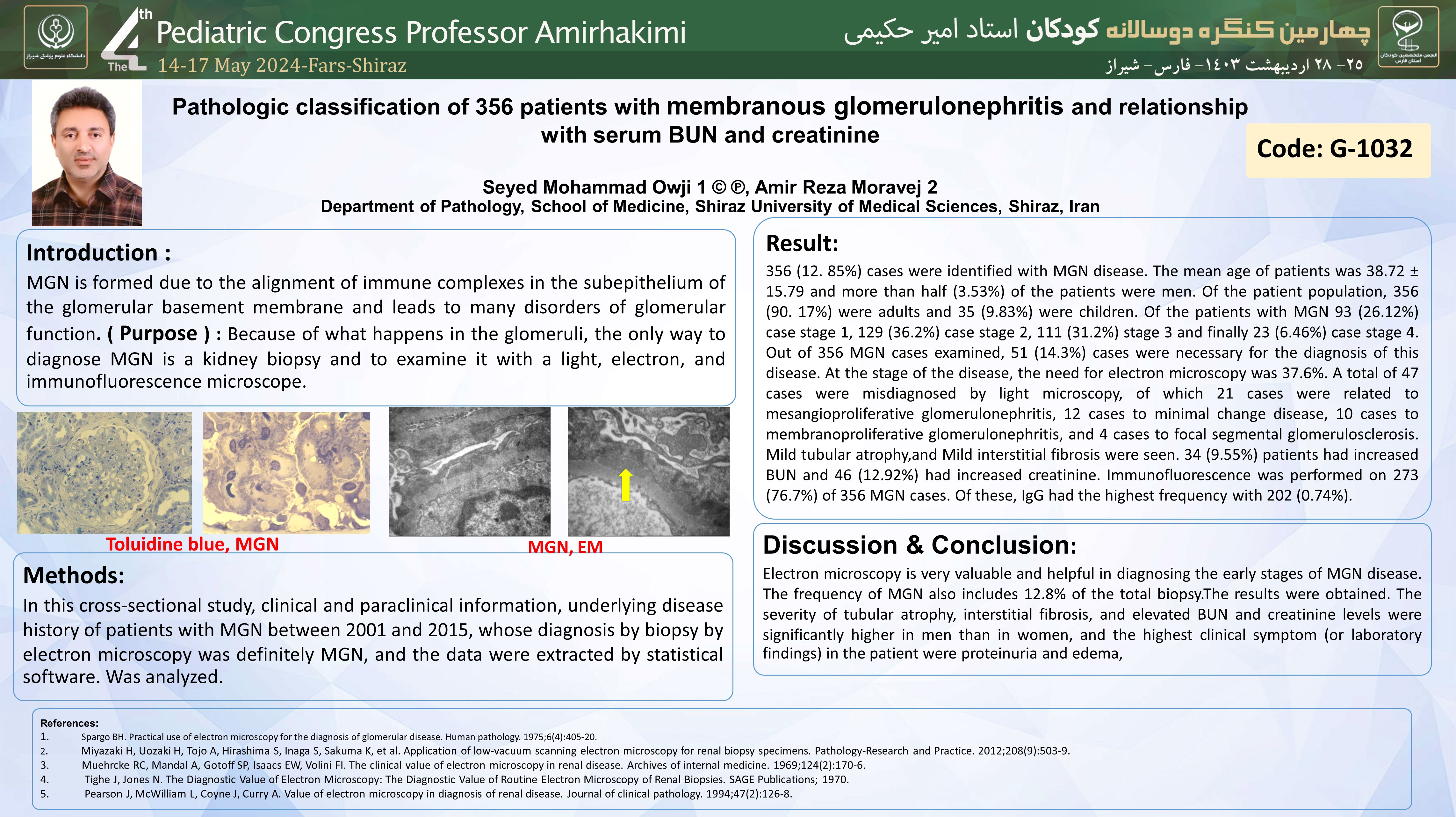
Background: MGN is formed due to the alignment of immune complexes in the subepithelium of the glomerular basement membrane and leads to many disorders of glomerular function. Because of what happens in the glomeruli, the only way to diagnose MGN is a kidney biopsy and to examine it with a light, electron, and immunofluorescence microscope. Method: In this cross-sectional study, clinical and paraclinical information, underlying disease history of patients with MGN between 2001 and 2015, whose diagnosis by biopsy by electron microscopy was definitely MGN, and the data were extracted by statistical software. Was analyzed. Results: 356 (12. 85%) cases were identified with MGN disease. The mean age of patients was 38.72 ± 15.79 and more than half (3.53%) of the patients were men. Of the patient population, 356 (90. 17%) were adults and 35 (9.83%) were children. Of the patients with MGN 93 (26.12%) case stage 1, 129 (36.2%) case stage 2, 111 (31.2%) stage 3 and finally 23 (6.46%) case stage 4. Out of 356 MGN cases examined, 51 (14.3%) cases were necessary for the diagnosis of this disease. At the stage of the disease, the need for electron microscopy was 37.6%. A total of 47 cases were misdiagnosed by light microscopy, of which 21 cases were related to mesangioproliferative glomerulonephritis, 12 cases to minimal change disease, 10 cases to membranoproliferative glomerulonephritis, and 4 cases to focal segmental glomerulosclerosis. Mild tubular atrophy,and Mild interstitial fibrosis were seen. 34 (9.55%) patients had increased BUN and 46 (12.92%) had increased creatinine. Immunofluorescence was performed on 273 (76.7%) of 356 MGN cases. Of these, IgG had the highest frequency with 202 (0.74%). Conclusion: Electron microscopy is very valuable and helpful in diagnosing the early stages of MGN disease. The frequency of MGN also includes 12.8% of the total biopsy.The results were obtained. The severity of tubular atrophy, interstitial fibrosis, and elevated BUN and creatinine levels were significantly higher in men than in women, and the highest clinical symptom (or laboratory findings) in the patient were proteinuria and edema,
